Understanding brain anatomy and function
Obtaining a general understanding of the brain and its functions is important to understanding the rehabilitation process.
Obtaining a general understanding of the brain and its functions is important to understanding the rehabilitation process.

There are three major parts of the brain:
The cerebrum is made up an outer layer, called the cortex, which is responsible for thinking, learning, memory, and emotions. Additionally, there are deeper, inner structures of the cerebrum, such as the thalamus, a small structure located slightly above the brainstem. This is the gateway for most of the sensory pathways. The thalamus plays a role in regulating awareness and emotional aspects of sensory experiences.
The brain stem begins underneath the brain and extends downward until it becomes part of the spinal cord. It is very important because it handles automatic functions like breathing, heart rate, and wakefulness.The reticular activating system is the part of the brain stem that responsible for wakefulness. This is a collection of neurons, located in the upper brain stem, that projects to and stimulates the areas of the cortex that is responsible for awareness, or the ability to think and perceive.
Consciousness requires both wakefulness and awareness. As you just learned, the parts of the brain that are responsible for wakefulness are located in a different area than the parts responsible for awareness. Disorders of consciousness are a result of either damage to one or both areas of the brain or damage to the connective network of neurons linking the areas responsible for wakefulness and awareness.
The brain receives blood flow with each beat of the heart. Major blood vessels connect the heart and brain. The blood carries vital nutrients, such as oxygen and glucose, to the brain.
Problems arise in the brain when blood flow becomes disrupted. It can be disrupted by either a clot (thrombus or embolus) that stops blood flow or when bleeding (hemorrhage) occurs. When there is bleeding, blood leaks out of the vessel and never makes it to the part of the brain it supplies. This could be caused from trauma or from other conditions such as aneurysm or stroke.
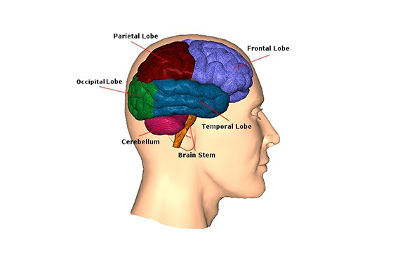
The frontal lobes are located in the front parts of the brain. They are over the eyes and behind a person’s forehead. There is a right frontal lobe and a left one.
Injuries to the frontal lobes may produce a variety of symptoms depending on the exact location of the problem. The frontal lobe contains the primary motor area that helps control movement and is responsible for higher-level problem-solving and reasoning skills.
A person may have one or more symptoms or different levels of severity of each symptom. The symptoms will depend on the extent and place of the injury.
The following symptoms may occur:
The temporal lobes are located on each side of the brain about where a person’s ears are found. There is a right temporal lobe and a left one.
The temporal lobes house the primary auditory area and are connected to key memory structures. Injuries to the temporal lobes may cause many symptoms, but most are related to understanding sounds or language and memory. The symptoms will depend on the extent and specific location of the injury.
The following symptoms may occur:
The parietal lobes are located on each side of the top areas of the brain. They sit behind the frontal lobes. There is a right parietal lobe and a left one. They house the primary sensory and visual or spatial area related to seeing how things fit together and construction.
Injuries to the parietal lobes may produce a variety of symptoms, but most are related to sensation or visual spatial skills. This relates to what a person feels on their skin, such as warmth or pain, or what a person recognizes about themselves. Symptoms will vary depending on the extent and specific location of the injury.
The following symptoms may occur:
The occipital lobe is in the back of the brain. Injuries to the occipital lobe may cause many symptoms related to vision. The symptoms will depend on the extent and location of the injury.
The following symptoms may occur:
The brainstem is like a tube that begins underneath the brain and extends down until it becomes part of the spinal cord. It is has three areas: the midbrain, pons, and medulla. The brainstem is very important because it handles automatic functions like breathing and heart rate.
The thalamus is a small structure located slightly above the brainstem. It is the gateway for most of the sensory pathways. The thalamus plays a role in regulating awareness and emotional aspects of sensory experiences, such as a reaction to fear or hunger. Symptoms of brainstem and/or thalamus injury depend on the extent and specific location of the
injury.
The following symptoms may occur:
Another condition that may occur when there are injuries near the brainstem and thalamus is storming, or hypothalamic instability or paroxysmal sympathetic storms. A storming episode may happen as a result of a stimulus or when the person is resting quietly. It may last several minutes and can happen at various times throughout the day. Signs of storming include an increase in heart rate, temperature and sweating. There also may be a rise in the blood pressure and muscle stiffness or rigidity during the episode. Storming happens because the injured brain is having difficulty controlling automatic bodily functions. Episodes of storming often decrease as the brain heals and the person becomes more responsive. Sometimes the doctor may order certain medicines to help decrease some of the symptoms.
The cerebellum is located in the back part of the brain underneath the cortex. It is under the occipital lobe and attached to the brainstem. Injuries to the cerebellum cause many symptoms related to balance and coordination. The symptoms will depend on the extent and specific location of the injury.
The following symptoms may occur:
